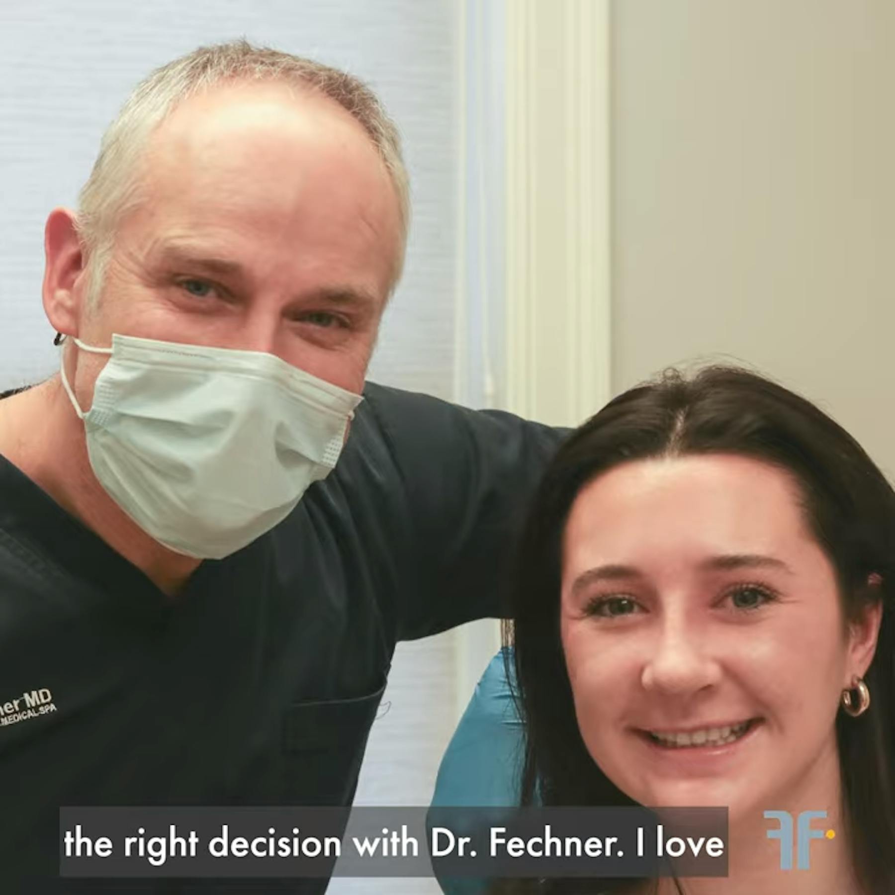
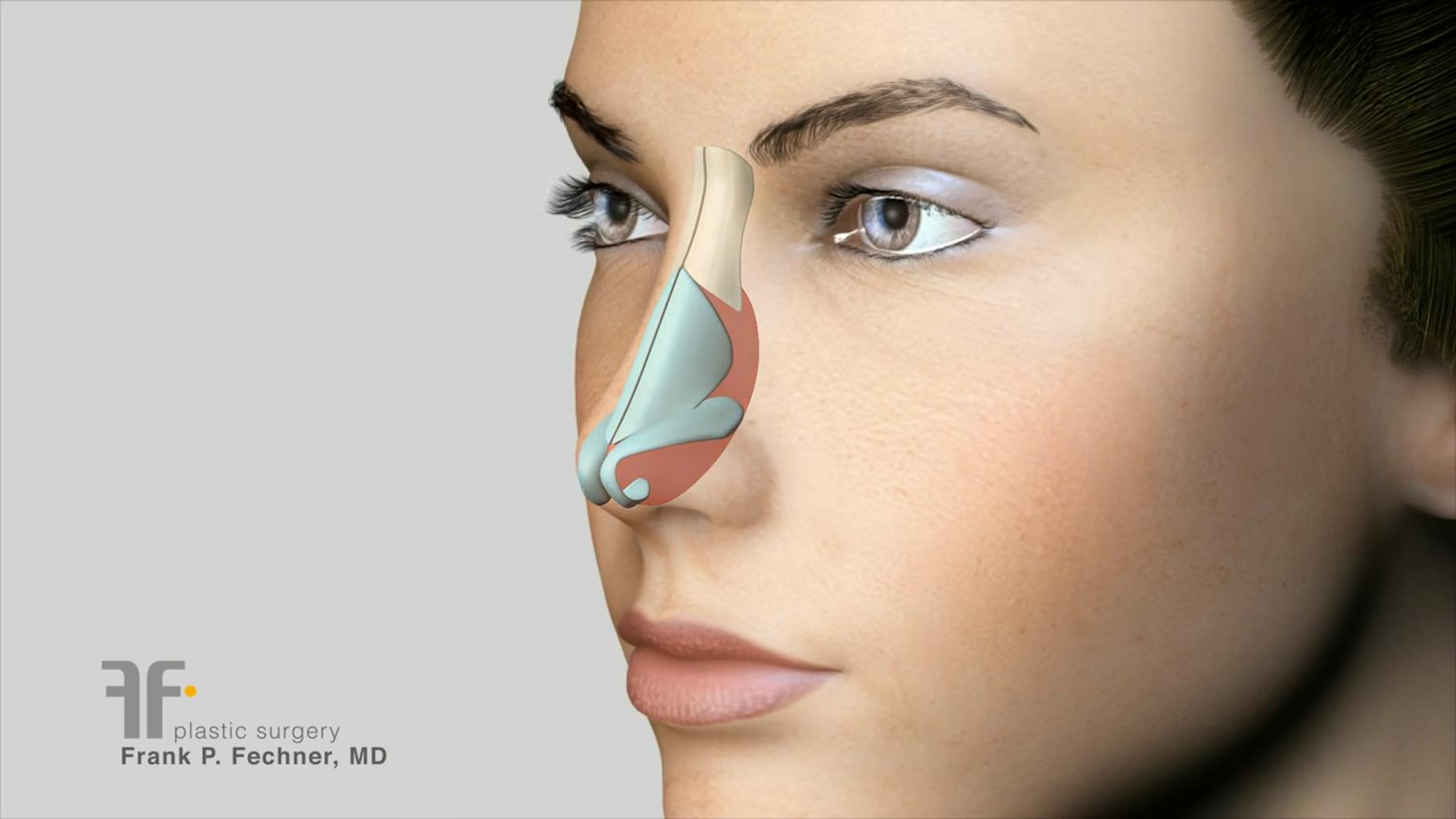
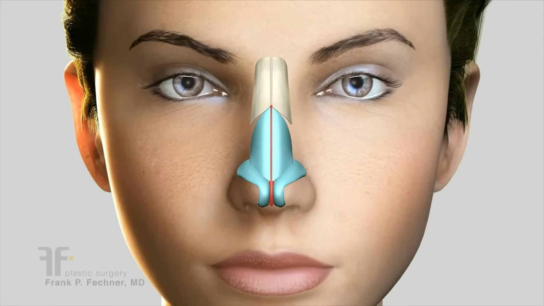
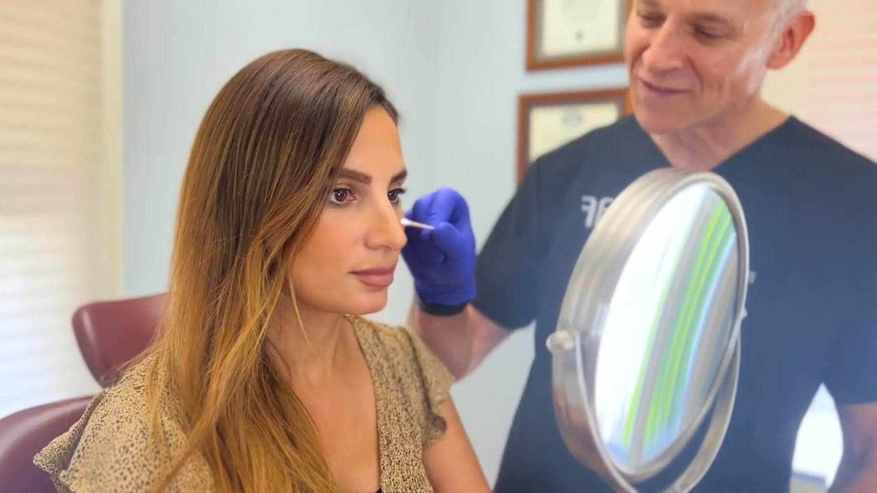
Rhinoplasty, commonly known as a "nose job," is a surgical procedure Dr. Fechner performs in Boston to alter the nose's shape, size, or structure for aesthetic or functional purposes.


Who Makes the Best Candidate for Rhinoplasty in Boston?
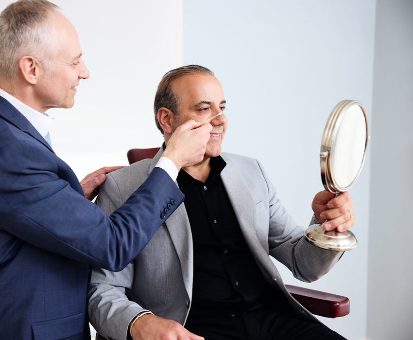
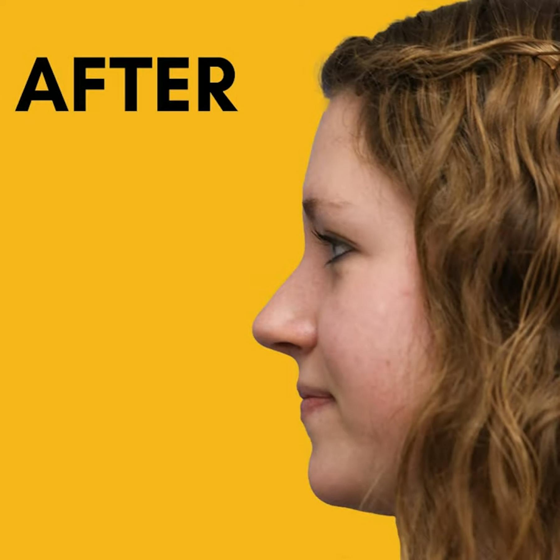
If you are unhappy with your nose for any reason, you deserve to obtain more information about a procedure that could help balance and harmonize your features. With rhinoplasty, the goal is to reshape the nose so that it doesn’t draw so much attention to itself. A rhinoplasty in Worcester is a great option for anyone seeking to cosmetically enhance their nose, provided the following attributes exist:
- Your nose is fully grown
- You are in good mental and physical health
- You are free from any major medical issues which might interfere with surgery
- You are realistic in your cosmetic goals
- You are a non-smoker, or willing to quit before and after surgery
- You are willing to follow Dr. Fechner's instructions both before and after your procedure